Table of Contents
- 1. Introduction
- 2. Language Acquisitions
- 3. Language Comprehension
- 4. fMRI/EEG Analysis Techniques
- 5. Tools for Neurolinguistic Computations
- 6. Experimental Findings and Brain Regions
- 7. Technical Details and Mathematical Formulations
- 8. Analysis Framework Case Study
- 9. Future Directions and Applications
- 10. Expert Analysis
- 11. References
1. Introduction
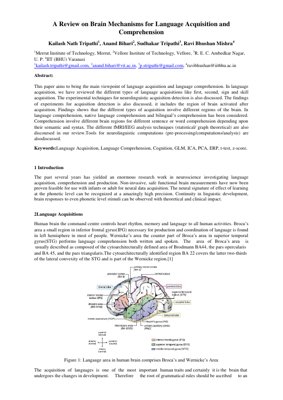
This paper reviews the main viewpoints on language acquisition and comprehension from a neurolinguistic perspective. It covers first, second, sign, and skill acquisition, along with experimental techniques such as fMRI and EEG. The neural signatures of learning at phonetic, lexical, and syntactic levels are examined, highlighting the roles of Broca's and Wernicke's areas.
2. Language Acquisitions
Language acquisition is a biologically determined process. The brain's Broca's area (BA44/45) and Wernicke's area (BA22) are central to production and comprehension, respectively. Acquisition involves distinct neural circuits depending on the type (L1, L2, sign).
2.1 First Language (L1) Acquisition
L1 acquisition occurs naturally during early childhood, progressing from babbling (6-8 months) to single words (10-12 months) and two-word stages (~2 years). Eric Lenneberg (1967) proposed a critical period ending at puberty, after which L1-like proficiency is rarely achieved. Neuroimaging shows that L1 processing relies heavily on left-hemisphere perisylvian regions.
2.2 Second Language (L2) Acquisition
L2 can be learned at any age, but proficiency rarely matches L1 if acquired after the sensitive period. fMRI studies reveal that L2 processing often involves additional recruitment of prefrontal and parietal regions, especially for late learners. The degree of activation in Broca's area correlates with proficiency.
2.3 Sign Language and Skill Acquisition
Sign language acquisition engages similar left-hemisphere language networks as spoken language, but also recruits visual-spatial areas. Skill acquisition (e.g., reading, writing) involves secondary neural pathways, often relying on the angular gyrus and occipito-temporal regions.
2.4 Neurolinguistic Experimental Techniques
Non-invasive techniques like fMRI, PET, and EEG are used to measure brain activity during language tasks. For infants, safe functional measurements are feasible. Event-related potentials (ERPs) and functional connectivity analyses provide insights into the temporal dynamics of acquisition.
3. Language Comprehension
Comprehension involves semantic and syntactic processing. Different brain regions are recruited depending on the complexity of sentences and words.
3.1 Native Language Comprehension
Native language comprehension primarily activates the left posterior superior temporal gyrus (STG, BA22) for phonological processing, and the left temporoparietal regions (angular gyrus) for lexico-semantic processing. Syntactic processing engages Broca's area.
3.2 Bilingual Comprehension
Bilinguals show overlapping but distinct neural networks for L1 and L2. L2 comprehension often requires greater activation in the left inferior frontal gyrus (IFG) and anterior cingulate cortex, reflecting increased cognitive control and effort.
4. fMRI/EEG Analysis Techniques
Statistical and graph theoretical methods are used to analyze neuroimaging data.
4.1 Statistical Methods (GLM, t-test, z-score)
The General Linear Model (GLM) is the standard for fMRI analysis, modeling BOLD signal as a linear combination of regressors. T-tests and z-scores are used for group-level inference. For EEG, ERP components (e.g., N400, P600) are analyzed using repeated-measures ANOVA.
4.2 Graph Theoretical Approaches
Graph theory models the brain as a network of nodes (regions) and edges (connections). Metrics like clustering coefficient, path length, and modularity reveal how language networks reorganize during acquisition and comprehension.
4.3 ICA and PCA
Independent Component Analysis (ICA) and Principal Component Analysis (PCA) are used for denoising and identifying latent neural sources. ICA separates mixed signals into independent components, while PCA reduces dimensionality.
5. Tools for Neurolinguistic Computations
Popular tools include SPM, FSL, AFNI for fMRI preprocessing and analysis; EEGLAB and FieldTrip for EEG; and custom scripts in MATLAB/Python for graph theoretical analysis. These tools enable preprocessing (motion correction, normalization), statistical modeling, and visualization.
6. Experimental Findings and Brain Regions
Key findings: L1 acquisition activates left perisylvian regions; L2 acquisition involves additional prefrontal and parietal areas. Comprehension of semantically anomalous sentences elicits an N400 ERP component, while syntactic violations elicit a P600. Bilinguals show reduced lateralization for L2.
7. Technical Details and Mathematical Formulations
The GLM for fMRI is expressed as: $Y = X\beta + \epsilon$, where $Y$ is the observed BOLD signal, $X$ is the design matrix, $\beta$ are parameter estimates, and $\epsilon$ is noise. For EEG, the ERP is computed as: $ERP(t) = \frac{1}{N}\sum_{i=1}^{N} x_i(t)$, where $x_i(t)$ is the $i$-th trial. Graph theory metrics: clustering coefficient $C = \frac{2E}{k(k-1)}$, where $E$ is the number of edges between $k$ nodes.
8. Analysis Framework Case Study
Case Study: L2 Acquisition in Late Learners
A group of 20 late L2 learners (age >12) underwent fMRI while performing a semantic judgment task in L2. Preprocessing: motion correction, slice-timing correction, normalization to MNI space. GLM analysis revealed significant activation in left IFG (BA44/45) and bilateral anterior cingulate. Graph theoretical analysis showed increased modularity in the fronto-parietal network compared to L1 controls. This indicates that late L2 acquisition relies on compensatory cognitive control mechanisms.
9. Future Directions and Applications
Future research should integrate multimodal imaging (fMRI+EEG) to capture both spatial and temporal dynamics. Machine learning models (e.g., deep learning) can predict language outcomes from brain connectivity patterns. Applications include early diagnosis of language disorders, personalized language learning interventions, and brain-computer interfaces for aphasia rehabilitation. The use of real-time neurofeedback could enhance L2 acquisition efficiency.
10. Expert Analysis
Core Insight: This review consolidates the neural basis of language acquisition and comprehension, emphasizing that different language types (L1, L2, sign) recruit partially distinct but overlapping brain networks. The critical period hypothesis remains a cornerstone, but recent evidence suggests that neural plasticity extends beyond puberty with appropriate training.
Logical Flow: The paper logically progresses from acquisition (types and techniques) to comprehension (native vs. bilingual), then to analysis methods and tools. The structure is clear, though the depth of experimental findings could be expanded.
Strengths & Flaws: Strengths include a comprehensive overview of key brain regions and experimental techniques. Weaknesses: the review lacks quantitative meta-analysis and does not address individual differences (e.g., genetic factors). The discussion of graph theory is superficial.
Actionable Insights: For researchers, integrating graph theory with machine learning can uncover predictive biomarkers for language proficiency. For educators, neurofeedback training targeting Broca's area may accelerate L2 learning. Clinicians can use ERP markers (N400, P600) for early detection of language impairments.
11. References
- Lenneberg, E. H. (1967). Biological Foundations of Language. Wiley.
- Friederici, A. D. (2011). The brain basis of language processing: from structure to function. Physiological Reviews, 91(4), 1357-1392.
- Hickok, G., & Poeppel, D. (2007). The cortical organization of speech processing. Nature Reviews Neuroscience, 8(5), 393-402.
- Ullman, M. T. (2001). A neurocognitive perspective on language: the declarative/procedural model. Nature Reviews Neuroscience, 2(10), 717-726.
- Perani, D., & Abutalebi, J. (2005). The neural basis of first and second language processing. Current Opinion in Neurobiology, 15(2), 202-206.
- Friston, K. J. (2011). Functional and effective connectivity: a review. Brain Connectivity, 1(1), 13-36.
- Luck, S. J. (2014). An Introduction to the Event-Related Potential Technique. MIT Press.
- Bullmore, E., & Sporns, O. (2009). Complex brain networks: graph theoretical analysis of structural and functional systems. Nature Reviews Neuroscience, 10(3), 186-198.